Aurora kinases

Aurora kinases: classification, functions and inhibitor design
The first protein kinase in a novel family of so-called Aurora kinases was detected during centromere cycle screening in fruit flies, also known as Drosophila melanogaster. Mutations in this protein caused development of monopolar spindles resembling the North Pole instead of centrosome uncoupling during cell cycle. That was the reason why the protein was given the name “Aurora” [1].
Since the discovery of Aurora kinases participation in mitosis cell division among different species this family of proteins became an object of interest for many researchers. It was discovered that overexpression and gene amplification of Aurora kinases is strongly associated with multiple cancers: gastric cancer, breast cancer, ovarian cancer and other tumors [1]. Because of that, members of this protein kinase family became novel therapeutic targets, and Aurora inhibitors were proven to effectively lower cancer activity.
Aurora libraries consist of small molecules that can potentially inhibit Aurora kinases. To conduct the design of inhibitors correctly, classification of this family of proteins should be considered. Nowadays, three known classes of Aurora kinases in mammal species can be identified:
-
Aurora kinase A (also known as Aurora A, AURKA, Aurora 2)
-
Aurora kinase B (also known as Aurora B, AURKB, Aurora 1)
-
Aurora kinase C (also known as Aurora C, AURKC, Aurora 3)
Let’s take a closer look at the structural aspects, role in cell processes and in cancer development of each class in the Aurora family.

Figure 1. Aurora kinase structural units [2].
Aurora A
Aurora A kinase has three distinct functional structural units or so-called domains: C-terminal unit and kinase unit, which are highly conservative among all the three classes, and N-terminal unit with a variety in length and little to no similarity between classes. Aurora A kinase structural unit, responsible for most of the Aurora kinase activity, has 71% of homology with Aurora B kinase unit and 60% of homology with Aurora C kinase unit. Phosphorylation of residue Thr288 leads to conformational changes which allow the protein to switch on its kinase activity [2].
Other conformational changes in protein include C-terminal and N-terminal domains folding to form the binding site of the protein. It is interesting to note that AURKA has specific hydrogen bonds between the adenosine and the hinge of the kinase structural unit, which can be used to develop Aurora A-specific inhibitors.
Regarding localization of AURKA in the cell, this protein is usually located near the centrosome. Aurora A is mainly active during the late G2 and late S phases of the cell cycle, and has its peak activity in mitosis [3]. There are several functions associated with the protein, which include: centrosome maturation, epigenetics, inhibition of tumor assembly, spindle assembly, mitotic entry regulation and so on.
Overexpression of Aurora A occurs in almost every kind of tumor - it was found in 29 of 33 cancer types. According to different studies, such overexpression causes genomic and chromosomal instabilities, an antiapoptotic effect, and epithelial-mesenchymal transition. It stimulates cell division even if DNA of cells is damaged, which results in overpopulation of abnormal cells in an organism [4].
Aurora B
Aurora B, being one of the members of Aurora family, also has three structural units, all of which except the N-terminal unit are homologous to units of Aurora A. N-terminal unit, on the other hand, is much shorter - the Aurora A N-terminal unit is nearly twice as long as the Aurora B N-terminal unit. The N-terminal unit of AURKB also contains a promoter to which transcription factors such as E2F1, E2F4, FOXM1 or DP2 can bind to regulate AURKB transcription [5]. Like AURKA, AURKB has a residue - Thr232 - which changes conformation of the protein after phosphorylation.
Cell localization of Aurora kinase B as a chromosomal passenger protein differs depending on the current phase of the cell cycle. Being transported by spindle and astral microtubules, it is also found in the cytoplasm and the equatorial cell cortex.

Figure 2. Aurora kinase B role in biological processes and in cancer development [5].
Functions of AURKB in normal cells and their role in cancer cells are presented in the figure above. It was discovered that Aurora B in tumors interacts with several other oncogenes and tumor suppressors, including Myc oncogenes, Bcr-Abl oncoprotein, cyclin K and p53-dependent tumor suppressor FBXW7.
Aurora C
Aurora C has a three-domain structure similar to Aurora A and Aurora B. However, because of alternative splicing its N-terminal domain exists as one of the three variants shown in the figure below.

Figure 3. Variants of AURKC [6].
Catalytic kinase structural unit of Aurora C has 60% homology with the Aurora A kinase structural unit, as it was stated before, and a pretty high 75% homology with the Aurora B kinase structural unit [2]. This may explain why both Aurora B and C interact with the same inner centromere protein (INCENP) and have similar functionality in biological processes.
Interestingly, while Aurora A and Aurora B are present in almost every cell of an organism, considerable expression of Aurora C proteins is found within cells going through meiosis (sperm and oocyte). AURKC function depends on the current localization and the phase of the cell cycle. Most known Aurora C kinase functions overlap with functions of Aurora B kinase [6].
Unfortunately, to this day little is known about the connection between cancer and Aurora C kinase functions, although high levels of Aurora C expression are detected in some tumors.
Aurora inhibitors
Most Aurora inhibitors were designed to inhibit Aurora A or Aurora B kinases. The reason for that is because there is much more information about their involvement in cancer development and their functions and conformations in general. Here we will review the most popular Aurora inhibitors, their targets and mechanism of action.
Table 1. Aurora inhibitors [5, 7] and their targets.
|
Aurora inhibitor |
Aurora A |
Aurora B |
Aurora C |
|
Barasertib (AZD1152) |
- |
+ |
- |
|
Alisertib (MLN8237) |
+ |
- |
- |
|
Hesperadin |
- |
+ |
- |
|
Danusertib (PHA-739358) |
+ |
+ |
+ |
|
PF-03814735 |
+ |
+ |
- |
|
AMG900 |
+ |
+ |
+ |
|
SP-96 |
- |
+ |
- |
|
GSK1070916 |
- |
+ |
+ |
|
AT9283 |
+ |
+ |
- |
|
CYC116 |
+ |
+ |
- |
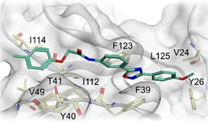
ChemDiv can offer Aurora A-B kinases targeted library containing 10000 compounds which demonstrate a similar binding mode compared to reported Aurora inhibitors.
References:
- Fu J, Bian M, Jiang Q, Zhang C. “Roles of Aurora kinases in mitosis and tumorigenesis”. Mol Cancer Res. 2007 Jan;5(1):1-10.
- Willems E, Dedobbeleer M, Digregorio M, Lombard A, Lumapat PN, Rogister B. “The functional diversity of Aurora kinases: a comprehensive review”. Cell Div. 2018 Sep 19;13:7.
- Naso FD, Boi D, Ascanelli C, Pamfil G, Lindon C, Paiardini A, Guarguaglini G. “Nuclear localisation of Aurora-A: its regulation and significance for Aurora-A functions in cancer”. Oncogene. 2021 Jun;40(23):3917-3928.
- Mou PK, Yang EJ, Shi C, Ren G, Tao S, Shim JS. “Aurora kinase A, a synthetic lethal target for precision cancer medicine”. Exp Mol Med. 2021 May;53(5):835-847.
- Borah NA, Reddy MM. “Aurora Kinase B Inhibition: A Potential Therapeutic Strategy for Cancer”. Molecules. 2021 Apr 1;26(7):1981.
- Quartuccio SM, Schindler K. “Functions of Aurora kinase C in meiosis and cancer”. Front Cell Dev Biol. 2015 Aug 20;3:50.
- Bavetsias V, Linardopoulos S. “Aurora Kinase Inhibitors: Current Status and Outlook”. Front Oncol. 2015 Dec 21;5:278.