PET imaging reveals synaptic loss in multiple sclerosis across preclinical and human studies
Click to view quick news summary (Spoiler)
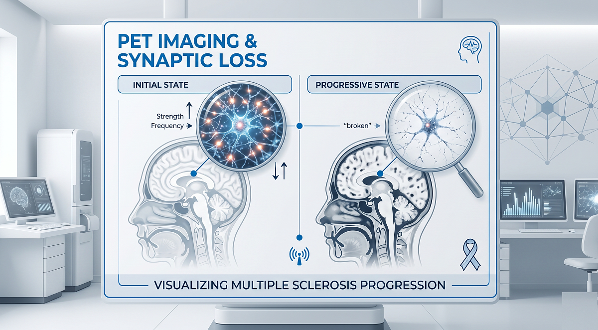
A novel SV2A PET imaging approach highlights extensive synaptic density reductions across subcortical, brain, and spinal cord areas in both multiple sclerosis models and living subjects, providing a sensitive baseline to monitor neurodegenerative pathology.
A new PET imaging approach that measures synaptic density in the spinal cord provides a quantitative way to assess the brain's functional wiring in patients with multiple sclerosis. With this personalized information, physicians can monitor disease progression and evaluate whether new treatments are working to protect or restore these critical connections. This research was presented at the Society of Nuclear Medicine and Molecular Imaging's 2026 Annual Meeting.
Multiple sclerosis is a condition that affects millions of people worldwide and can cause physical disability, fatigue, and cognitive impairment. While multiple sclerosis is traditionally viewed as a disease that damages the protective coating of nerves, there is also another, more subtle type of damage: the loss of synapses, which are the vital connection points where brain cells communicate.
Quantifying Spinal Cord Synaptic Density
“Although the spinal cord is a primary and often early site of inflammatory and neurodegenerative pathology in multiple sclerosis, in vivo quantification of synaptic density in this region has not yet been explored,” said Pou Hong Justin Chia, a graduate student at the Centre for Addiction and Mental Health at the University of Toronto, the lead presenter of the study. “To address this knowledge gap, my colleagues and I investigated a specialized imaging technique called SV2A PET to visualize and quantify this loss of connections in the spinal cord of a mouse model of multiple sclerosis and, crucially, in the brains and spinal cords of living multiple sclerosis patients.”
In the study, researchers conducted 18F-SynVesT-1 PET scans in mice with experimental autoimmune encephalomyelitis, a widely used mouse model of multiple sclerosis, and in healthy control mice. Spinal cord regions of interest were defined, and total volume of distribution and radiotracer binding were quantified and compared between groups. To provide a translational context, 11C-UCB-J PET imaging was performed on six multiple sclerosis patients and six healthy controls in collaboration with Yale University. Total volume of distribution maps were generated and human PET data were compared.
Preclinical Correlation and Human Clinical Trial Data
In the multiple sclerosis-model mice, 18F-SynVesT-1 PET successfully detected significant reductions in synaptic density within specific regions of the spinal cord, which were corroborated by binding studies. In the human PET study, multiple sclerosis patients exhibited a 16.4 percent reduction in 11C-UCB-J binding across the brain as compared to healthy controls. Widespread reductions were also observed in subcortical and spinal cord regions, mirroring the extensive synaptic pathology seen in the preclinical model.
According to Chia, this research provides direct evidence in living subjects that synaptic loss is a widespread feature of multiple sclerosis. “Understanding how and where these connections are lost can help explain the symptoms patients experience and give doctors and researchers a more sensitive way to detect disease-related changes, monitor progression over time, and better understand how multiple sclerosis and other neurological diseases affect the brain and spinal cord,” he said.
Currently, SV2A PET imaging in multiple sclerosis is available for clinical trials at specialized academic centers. While it is not yet a routine part of standard clinical care, the data from this pilot study is a necessary step toward larger clinical trials. If validated in larger studies, this imaging approach could be integrated into clinical practice and drug development over the next several years.